Scientists successfully used stem cells from human amniotic fluid to boost the growth of robust, functional blood
vessels in healing hydrogels injected into mice.
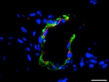
Two weeks after implantation, hydrogel seeded with amniotic fluid stem cells showed growth of mature blood vessels.
The picture shows cell nuclei (blue), endothelial cells (red) and smooth muscle cells (green). The scale bar is 20
microns.
Image credit: Jacot Lab/Rice University
The team – led by Jeffrey G. Jacot, an associate professor of bioengineering at Rice University and Texas
Children’s Hospital in Houston, TX – report
their work in the Journal of Biomedical Materials Research Part A.
The researchers are working on ways to use cells taken from the amniotic fluid of pregnant women to help heal babies
born with heart defects.
Amniotic fluid is often drawn in standard tests done during pregnancy. The fluid is usually discarded once the tests are
complete, but the team explains it is now showing promise as a source of implants made from a baby’s own genetically matched material.
Prof. Jacot says the stem cells in amniotic fluid are valuable because they can differentiate into many other types of
cell – including endothelial cells that make blood vessels.
He comments on their achievement:
“The main thing we’ve figured out is how to get a vascularized device: laboratory-grown tissue that is made
entirely from amniotic fluid cells. We showed it’s possible to use only cells derived from amniotic fluid.”
Healing hydrogels are increasingly being developed for use in medicine. For example, they can be used as 3D “scaffolds” in
tissue engineering, and they can also be used to target the delivery of drugs precisely to where they are needed.
Hydrogel scaffolds made with a biopolymer called fibrin are widely used in wound healing and promoting angiogenesis –
where new blood vessels branch off from ones already present in neighboring tissue.
Fibrin hydrogel with amniotic cells grew more robust blood vessels
For their study, the team used mice to investigate what happens when you combine the angiogenesis properties of fibrin
hydrogel with the endothelial cell generating effect of amniotic stem cells.
They first made some modifications to the fibrin hydrogel, which on its own was too stiff and degraded too quickly. Adding
polyethylene glycol to the fibrin made the hydrogel much more pliable and robust.
To encourage the amniotic stem cells to differentiate into endothelial cells, the researchers treated them with vascular
endothelial growth factor.
The researchers found that mice injected only with modified hydrogel developed thin fibril structures typical of
angiogenesis, but the mice injected with hydrogel seeded with amniotic cells showed much more robust new blood vessel
networks.
Similar results using hydrogel seeded with mesenchymal cells taken from bone marrow are possible, but this method does not
guarantee a tissue match, Prof. Jacot explains.
The authors also experimented using hydrogel seeded with endothelial cells but this did not work as well as they expected,
he adds.
The team is now looking at how amniotic cells might be used to make biocompatible patches to repair hearts
of infants born with birth defects and also for other procedures.
Funds for the study came from the National Institutes of Health and the National Science Foundation.
In February 2015, Medical News Today reported an MIT-led study where scientists created an injectable hydrogel that delivers drugs in a timed fashion. Writing in Nature
Communications, the team describes that not only can the hydrogel offer targeted drug delivery, each gel component can
be modified to deliver different drugs at different rates, offering the possibility of tailoring it to individual patient
needs.