Remember the game Operation? You need to carefully remove the body part without nicking the sides or the buzzer will sound.
Only imagine pulling out the funny bone when it’s surrounded by vessels controlling urinary and sexual function. That’s the issue when treating prostate cancer.

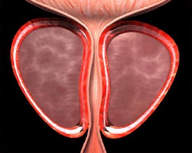
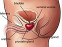
The prostate is a tiny organ surrounded by critical structures. When radiation treatments deliver dose to any of these structures, it can sometimes lead to problems such as erectile dysfunction and bladder or rectal irritation.
“We always have to keep cure as our first priority, but quality of life is a major secondary concern for men with prostate cancer,” says Patrick W. McLaughlin, M.D., professor of radiation oncology at the University of Michigan Medical School.
“In the past cure came at a steep price in lost quality of life, but with modern refinements it is increasingly possible to meet the new standard of successful prostate cancer treatment: cure with quality of life.”
McLaughlin is the senior author of a paper published in Lancet Oncology that looks at how MRI and a clear understanding of the functional anatomy (and its variations from patient to patient) can allow radiation oncologists to plan a course of treatment that spares these critical structures.
The team started by defining the critical functions and structures that run through or near the prostate. These include the nerves, vessels and sphincters that control bladder function, erectile function and rectal function.
“The benefit of the functional anatomy approach goes well beyond improving sexual function. It has improved urinary and rectal function as well,” McLaughlin says.
Both radiation oncologists and surgeons are conducting ongoing research to improve their understanding of the functional anatomy. This is critical for both disciplines as they work to eliminate cancer while preserving function.
“We argue that current rates of side effects and changes after treatment are not fixed, and that further potential improvements in function preservation are possible and likely by pursuing this vessel-sparing approach,” says co-author Jae Lee, M.D., Ph.D., a radiation oncology resident.
The study authors also found that MRI was a critical tool for accurately outlining the prostate anatomy and planning radiation therapy. They could easily and precisely see the borders of the prostate on MRI. Images from CT are much less clear, and it’s common to overestimate the area that needs to be treated. With a clear outline of the prostate and other critical structures on MRI, radiation oncologists can precisely target treatment to the prostate while avoiding these critical erectile tissues. The technique is called vessel-sparing radiation.
Aggressive prostate cancer is often treated with a combination of implanted radiated seeds and external beam radiation. The combination is more effective than either method alone in treating aggressive cancers.
Of 49 patients treated with combination seeds plus external beam radiation with at least five years of follow-up, 92 percent reported they were still able to be sexually active.
“We found no difference in quality of life for men given aggressive treatment. If you define the functional structures and limit dose to them, you can achieve cure and excellent quality of life,” says co-author Daniel E. Spratt, M.D., chief of the genitourinary radiation oncology program at the University of Michigan.
Vessel-sparing radiation requires physicians train in MRI anatomy to recognize and identify key structures. McLaughlin has previously developed a tool that is available free online called Prostadoodle. It includes a section on defining the erectile vessels.
In addition, MRI could be helpful in guiding patients to the best treatment option based on their unique anatomy. Is the tumor outside of the prostate gland? That would suggest the need for radiation therapy after surgery, if surgery is chosen as primary treatment. Does the patient have a short urinary sphincter? While rare, these patients have a higher risk of incontinence with surgery.
“For patients who appear to have slow-growing, non-aggressive cancers, MRI can confirm there is no aggressive cancer present. For such patients, surveillance is an excellent choice,” McLaughlin says. “By avoiding treatment altogether when appropriate, all the side effects and quality of life impact from treatment is avoided.”
“On the other end of the spectrum, MRI may actually reveal more serious cancers not sampled by the biopsy. This can shift treatment to a more aggressive approach necessary to cure such cancers.”
Related posts:
 Results From STRIVE Trial Of Enzalutamide Versus Bicalutamide In CRPC Published In Journal Of Clinical Oncology
 San Francisco sheds light on a new mode of drug resistance to emerging therapies in metastatic prostate cancer
 Researchers Launch Clinical Trials to Test Novel Cellular-immunotherapy to Treat Three Types of Blood Cancer
 Male Friends not to be Missed Osteoporosis Screening