What is Gallbladder Cancer?
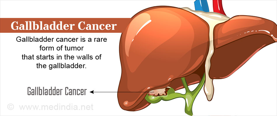
Gallbladder cancer is a rare cancer in which malignant (cancerous) transformation of cells occur in the wall of the gallbladder.
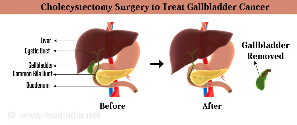
The gallbladder is a pear-shaped organ under the liver that stores bile. Bile is a fluid produced by the liver to digest fats. When food is being digested in the stomach and intestines, the gall bladder contracts and releases the bile into a tube called the cystic duct. The cystic duct joins with a duct from the liver called the common hepatic duct and together they form the common bile duct. The common bile duct then joins the pancreatic duct to empty the contents into the small intestine to aid the digestive process.

Gall bladder cancer is more commonly seen in South America, Central and Eastern Europe, Japan and Northern India. Certain ethnic groups e.g. native American Indians and Hispanics appear to be prone to gallbladder cancer. Women are affected more commonly than men. Cure is possible if the diagnosis is done early enough. Unfortunately a diagnosis is often made once symptoms occur, i.e. after the cancer advances. In such cases, the outlook is poor. Surgery is the most effective treatment for gallbladder cancer. Radiation and chemotherapy are also used.
Statistics on Gall Bladder Cancer
Gallbladder cancer:
- Is the most common cancer of the biliary tract (80 -90%) worldwide according to autopsy studies
- Has high rates of incidence in South America (Chile and Bolivia) and Asia (South Korea) and the lowest in Africa.
- Is seen up to 65% in less developed countries.
What are the Types of Gallbladder Cancer?
- Adenocarcinomas – These cancers start in the gland-like cells of the gallbladder and account for 9 out of the 10 gallbladder cancers. Among adenocarcinomas, papillary adenocarcinoma or papillary cancer accounts for 6% of gall bladder cancers.It has cancerous cells arranged as finger-like projections. Papillary adenocarcinoma usually does not spread to the liver or lymph nodes and has a good survival rate.
- Other rarer types are adenosquamous carcinoma, squamous cell carcinoma, small cell carcinoma and sarcoma.
What are the Stages of Gallbladder Cancer?
Staging is an important part of cancer management.
The stages of gallbladder cancer are grouped according to 3 main criteria:
- The size and spread of the tumor inside the layers of the gallbladder
- The spread of the tumor to the lymph nodes
- The spread of the tumor to a different part or organ of the body
Gallbladder cancer may be
- Localized (stages 1 and II) where the cancer is contained within the walls of the gallbladder. Surgery can be performed to remove the cancerous growth or the gallbladder itself.
- Advanced (stages III and IV) when the cancer has spread to the tissues outside the gallbladder either to the lymph nodes, blood vessels or other organs. The entire cancer cannot be removed or is not resectable except in the case of some stage 3 cancers. Stage 4 cancers can reappear either in the gallbladder or other organs after the first treatment.
What are the Causes and Risk factors of Gallbladder Cancer?
The cause of gall bladder cancer and the molecular mechanism by which the cancer develops still remain unclear. Risk factors for gallbladder cancer include the following:
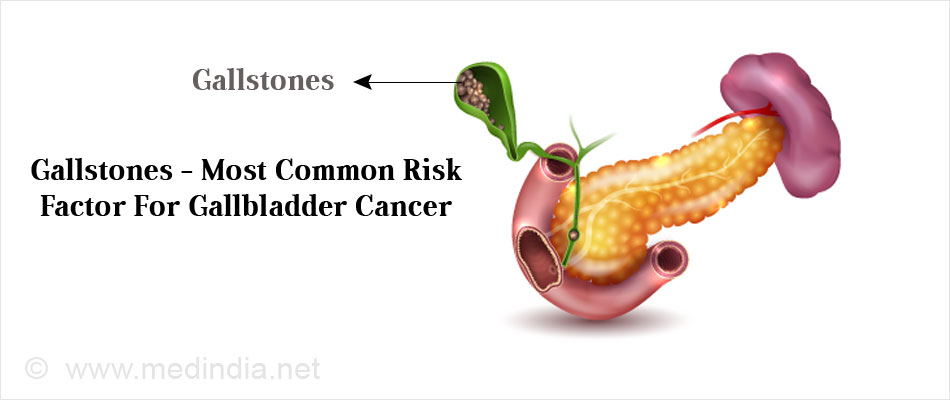
- Presence of gallstones: Gallbladder Stones are found in 85% of the people diagnosed with the cancer. The association increases with the size of the stones. The gallbladder also releases bile more slowly when gallstones are present exposing the cells to chemicals in bile that could cause irritation and inflammation, thereby predisposing to cancer.

- Chronic inflammation: Recurrent and chronic inflammation of the gallbladder could also cause changes in the DNA of the cells making them grow abnormally and resulting in cancerous transformation. Inflammation can also lead to calcification of the gallbladder. This condition is called ‘porcelain gallbladder’. People with porcelain gallbladder are at a high risk developing the cancer.
- Pancreatico-biliary duct junction anomalies: Abnormalities at the confluence of the pancreatic and the bile ducts known as the pancreaticobiliary duct junction might cause a reflux of the pancreatic juices back into the gallbladder and the bile ducts causing inflammation. This mechanism is a more common cause of gallbladder cancer in the Asian populations.
Other risk factors of gallbladder cancer include:
- Gender: Gallbladder cancer is twice more common in women than men.
- Age Gallbladder cancer is usually noted in older individuals
- Obesity: Obesity increases the risk of getting the cancer
- Ethnic groups: Ethnic groups like Native American Indians and Hispanics are at a high risk
- Chronic bacterial infections of the bile ducts
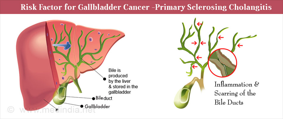
- Primary sclerosing cholangitis – Primary sclerosing cholangitis is the inflammation and fibrosis of the bile ducts inside and outside the liver

- Gallbladder polyps or growths in the walls of the organ
- Cysts of the common bile duct
What are the Symptoms and Signs of Gallbladder Cancer?
The early stages of gallbladder cancer may not produce any noticeable signs or symptoms.
Gallbladder cancer symptoms and signs include:
- Jaundice, where yellowing of the skin and whites of the eyes occurs

- Pain in the upper right part of the abdomen
- Fever
- Nausea and vomiting
- Lumps in the abdomen due to an enlarged gallbladder and/or spread of the cancer to other areas like the liver
- Rarer symptoms are loss of appetite accompanied by weight loss, bloating, itchy skin, dark urine and grey colored stools.
How is Gallbladder Cancer Diagnosed?
An early diagnosis of gallbladder cancer is difficult since the early stages of the cancer go unnoticed due to the lack of specific signs or symptoms. The clinical features produced by gallbladder are mimicked by a number of other conditions. Early symptoms mime gallbladder inflammation due to gallstones. The later symptoms are similar to those produced by biliary and stomach obstruction.
The cancer may be an incidental finding when the gallbladder is removed for other reasons or if an ultrasound done for some other reason shows a mass in the gallbladder.
Diagnosis begins with elicitation of history and physical examination. Once suspected, blood tests and imaging techniques are used to diagnose gallbladder cancer.
Blood tests include the following:
- Liver function tests are performed to know if the liver has been affected by the gallbladder cancer
- Levels of Carcinoembryonic Antigen (CEA) are checked for. CEA is a protein produced by both cancer cells and normal cells. Higher levels of CEA may indicate gallbladder cancer or other conditions
- Another similar test is CA 19-9 assay. CEA and CA 19-9 are referred to as ‘tumor markers’
Imaging studies can include:
- Abdominal ultrasound to visualize the gallbladder and other abdominal structures. It can be done with a probe applied on the abdomen, through an endoscope introduced through the mouth, or through a small incision in the abdomen (laparoscopic ultrasound).

- Computed Topography or a CT scan which takes a series of X-ray pictures to determine the stage of the cancer and whether it has spread to the liver and lymph nodes. An oral contrast liquid is given to the patient prior to the scan to outline the structures inside the body. A CT-angiogram can determine if the cancer has spread to the neighboring blood vessels.
- Magnetic Resonance Imaging or an MRI scan uses radio waves and strong magnets to provide accurate images of soft tissues inside the body. It provides more details than a CT scan.
- Cholangiography is an imaging test that aids in visualizing the duct system that carries bile and helps to see if there is a tumor blocking it. It also helps the surgeon to plan surgery for the cancer. The endoscopic retrograde cholangiopancreatography or ERCP uses an endoscope that is sent down the throat. It is invasive but helps to take a sample of the cells to examine under a microscope. In the percutaneous transhepatic cholangiography or PTC, a dye is injected through a needle through the skin prior to an x-ray to detect any block in the bile ducts
- Chest X-Ray
- Laparoscopy aids in direct visualization of organs and obtaining a biopsy
What is the Treatment and Management of Gallbladder Cancer?
Surgical removal is the most effective treatment of gallbladder cancer. A completely curative resection is however possible only in a minority of patients. This is because most patients seek treatment in an advanced stage of the cancer when surgery may not be possible.
When possible, the gallbladder and nearby tissues (including lymph nodes) are removed. Surgery is associated with significant mortality and morbidity.
The surgery can be a:
- Simple cholecystectomy (done laparoscopically or as an open operation) to remove the gallbladder alone

- Extended cholecystectomy that involves removing the gallbladder and parts of organs and / or lymph nodes that the cancer may have spread to.
In cases where operative removal is not possible, alternatives are called in:
- Surgical biliary bypass: A new pathway for drainage of bile is created
- Endoscopic stent placement: A stent (thin, flexible tube) is used to drain the bile into the small intestine
- Percutaneous transhepatic biliary drainage: This is also a procedure for draining bile
These procedures relieve the patient of jaundice.
Apart from surgery, radiation therapy and chemotherapy are also available for the treatment of the cancer.
Radiation Therapy
Radiation therapy is a cancer treatment where high-energy x-rays or other types of radiation are used to kill cancer cells or prevent them from growing. External beam radiation is used to treat gallbladder cancer. It may be given after surgery to destroy remaining cancer cells or in non-resectable cancers. In external radiation therapy, a machine outside the body is used to send radiation toward the cancer. The total dose of radiation therapy is sometimes divided into several smaller, equal doses which are delivered over a period of time spanning over several days.
Chemotherapy
Chemotherapy drugs may be used to stop the growth of cancer cells, either by killing the cells or by preventing them from dividing.
Chemotherapy drugs used to treat gallbladder cancer are:
- Gemcitabine
- Cisplatin
- 5-fluorouracil
- Capecitabine
- Oxaliplatin

What is the Prognosis for Gallbladder Cancer?
The outlook (prognosis) and treatment options for gallbladder cancer depend on a number of factors.
- The stage of the cancer: The extent of the cancer plays a vital deciding role
- Whether the cancer was diagnosed early
- Whether it is a recurrent case. A recurrent gallbladder cancer is one that has reappeared after complete treatment
- Whether complete surgical removal is possible
- The type of gallbladder cancer (based on microscopy)
- The general health of the patient
Localized cancer can be completely removed surgically and has a good prognosis. Advanced stages or recurrent cases have a poor prognosis. The median survival for advanced disease is short (2-4 months).